
The vacuum bell is a natural, non-surgical method for correcting pectus excavatum (chest wall depression)

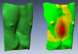
The causes of pectus excavatum (PE) are unknown. It is likely that the deformity is genetically influenced and occurs in about 35% of cases within families. Doctors often observe abnormal growth of the costal cartilage, which is believed to be related to the development of the deformity. It has also been noted that the severity of the deformation is influenced by the diaphragmatic sternal (chest) attachment. The extent of the deformity depends on the degree of its dysplasia (immaturity). Based on the above, physical activities that increase the frequency and volume of breathing contribute to the enlargement of the deformity in children and adolescents. However, there is no scientific evidence to support this theory.
Due to pathological and sometimes asymmetric growth of the ribs in the anterior chest wall, both minor and significant deformities can develop. Currently, specific genetic markers for PE have not been identified. However, genetic or familial predisposition is found in 35% of cases. It is worth noting that PE often coexists with various genetic conditions, such as Marfan syndrome, Ehlers-Danlos syndrome, and Poland syndrome.
According to various sources, PE occurs in 1 in 300 to 400 newborns, with a predominance in males (male-to-female ratio of 3:1). PE accounts for approximately 90% of all congenital chest deformities.
The impact of chest deformity on the development and functioning of internal organs remains controversial. It has been noted that most PE patients in childhood and adolescence are more prone to respiratory infections, often complicated by pneumonia. From a functional perspective, PE is asymptomatic in many patients. In some cases, patients experience increased fatigue, discomfort, palpitations, especially during exertion, shortness of breath, and chest pain. Cardiac function is generally not impaired, but 20-60% of patients may have mitral valve prolapse. Echocardiography typically shows some degree of ventricular compression and heart displacement, and in rare cases, regurgitation of the mitral or tricuspid valve. Patients with PE who are 40 years of age and older may develop angina pectoris and varying degrees of congestive heart failure, which are poorly responsive to treatment. Typically, after surgical treatment at a young age, echocardiographic analysis shows normalization of cardiac function, with an improvement in cardiac index during physical exertion.
PE is often noticed at birth or in the first year of a child's life. During growth, the deformity typically progresses. The peak progression of the deformity occurs during the period of sexual maturation when there is rapid growth of the entire body. During this period, the deformity can rapidly progress over 6-12 months. It is during adolescence (between the ages of 10 and 16) that most patients require surgical treatment.


 Customer support
Comments area
No comments yet