
The vacuum bell is a natural, non-surgical method for correcting pectus excavatum (chest wall depression)


Functional disorders refer to disruptions that hinder the normal functioning of the body. For example, pectus excavatum deformation (PED) leads to a reduction in chest volume, which in turn affects the essential functions of internal organs. Some patients with PED report chest pain or discomfort in the thoracic spine. The cause of these pains is unknown, but it is presumed that they arise due to postural issues frequently observed in patients with PED.
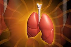
Symptomatic deterioration in lung function among PED patients manifests as a decrease in TLC (total lung capacity) and disruptions in breathing mechanics. The onset of these issues is linked to a decrease in intrathoracic volume. Scientifically, it has been proven that breathlessness during physical exertion in PED patients primarily occurs due to a reduction in lung reserve. Physicians note that most PED patients typically complain of breathing problems during adolescence or early adulthood. Breathlessness is not characteristic of patients under the age of 10.
In 1984, J. Cahill reported that after surgical reconstruction of PED, there was a slight increase in TLC, but maximal lung ventilation improved. Physical endurance also increased in terms of the duration of possible exercise and VO2max (maximum oxygen consumption).
After surgical treatment, patients exhibited a decrease in heart rate (HR) at a given level of physical exertion, with no change in VO2max to maintain work efficiency. The observed reduction in HR during similar exertion can be attributed to an increase in stroke volume due to an expanded intrathoracic space.
In 1967, Weg and colleagues conducted a comparative analysis of 25 American Air Force recruits with PED and healthy trainees. Despite similar lung measurements and TLC values in both groups, maximal lung ventilation (MLV) results showed significant differences, with PED patients having significantly lower values.
In 1996, P. Quigley conducted a study of 36 adolescents (average age 16 years) with PED. According to the authors, there was a significant reduction in forced vital capacity in the group of patients with chest deformity compared to a control group of peers without deformity. Additionally, they noted a recovery of this parameter following reconstructive surgery to correct the deformity.

The displacement of the sternum and ribs backward can lead to compression of the right ventricle of the heart. Earlier pathological-anatomical studies have confirmed this phenomenon in PED patients. Angiographic examinations of the heart's blood vessels revealed impaired coronary artery function. Research conducted by R. Peterson on the effects of workload on the cardiovascular system in 13 PED patients showed increased endurance after surgical deformity reconstruction and reduced recovery times.
After the age of 40, PED patients experience a sharp increase in the development of congestive heart failure and angina pectoris. This condition is difficult to treat, so many cardiologists recommend undergoing this operation.


 Customer support
Comments area
No comments yet